

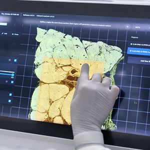
Scientific Operations, Spatial Team
The application of spatial genomics is unravelling biological architecture, expanding our knowledge of tissue structure and cellular interactions, and enabling detailed investigations into disease and development.
Through our work and knowledge we contribute to the emergence and onboarding of quality spatial technologies to support the science at the Sanger Institute. Our work aims to generate data that enhances our understanding of the human body, improving how we recognise, diagnose, and treat diseases. We also engage with the public to highlight how our efforts support scientific progress in society.
Current Spatial Technologies
Xenium in situ: V1 and Xenium Prime
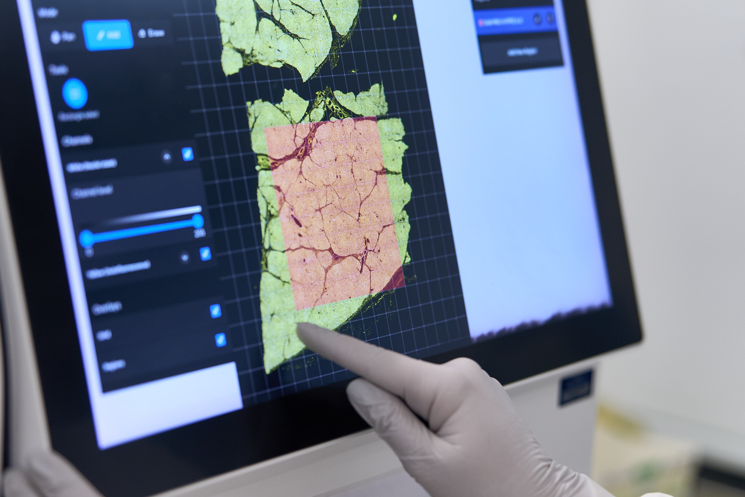
The Xenium platform delivers high-plex in situ at subcellular resolution with nanometer precision and offers a complete solution, including comprehensive panel offerings, to achieve fast and robust single cell spatial insights.
The Spatial Team have 5 Xenium machines available and are able to take your sample all the way from sample processing to ready to analyse data.
Our Xenium workflows are compatible with FFPE, Fresh Frozen and Fixed Frozen tissues from human and mouse samples. Custom gene panels and protein panels can also be incorporated.
Visium Spatial: Visium HD WT Panel and Visium HD 3′
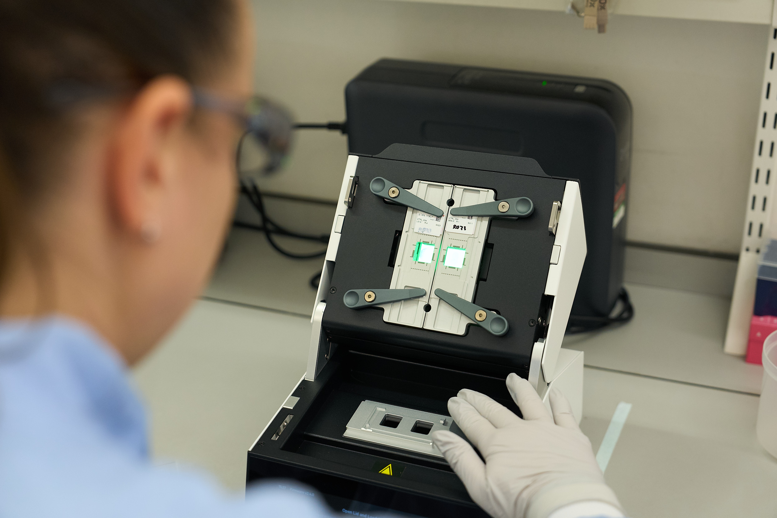
The Visium platform delivers unbiased, whole transcriptome spatial gene expression analysis at single cell scale with unmatched spatial data quality and offers a flexible assay portfolio to study a broad range of tissues and species.
The Visium HD WT (whole transcriptome) Panel is compatible with FFPE and Fresh Frozen tissues from human, mouse or rat samples.
The Visium HD 3′ assay offers de-novo whole transcriptome data and has the potential to be used with fresh frozen samples of any species.
Future Spatial Technologies
Atera in situ will be coming to Scientific Operations, Spatial Team Autumn 2026.
Sample Processing & Histology
The Spatial Team supports the end-to-end workflow for our Spatial Technologies. We can provide:
Sample Processing
This includes the processing of tissue samples into either formalin-fixed paraffin-embedded (FFPE) blocks, fresh frozen (FF) OCT embedded blocks or fixed frozen OCT embedded blocks.
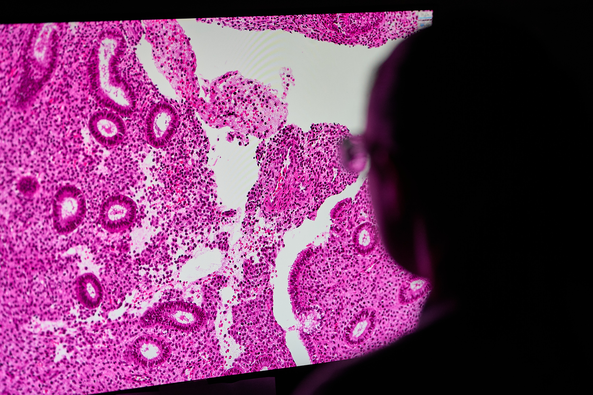
Sectioning
This includes taking sections for initial morphology checks, RNA quality checks, and sectioning for use in our spatial workflows, with the ability to capture multiple samples within these limited capture areas.
Quality Control H&E staining and imaging
We always recommend a morphology check in the form of a Haematoxylin and Eosin (H&E) stain, to be carried out on all samples before proceeding with any spatial omics workflow. H&E staining enables tissue morphology to be easily visualised. We use this as our tissue quality control and also to identify any regions of interest for spatial omics analysis.
Molecular QC
To ensure the samples are of sufficient molecular quality for the Spatial Omics technologies, we can also provide RNA quality checks, reporting back either DV200 (for FFPE samples) or RIN (for FF samples) score of the samples. This is especially important for the Visium HD 3’ workflow.
Sanger people

Dr Katy Tudor
Technical Specialist

Dr Harriet F Johnson
Advanced Research Assistant

Jasmine Halliwell
Advanced Research Assistant

Holly Anderson
Advanced Research Assistant
Previous Sanger people

Ilaria Mulas
Advanced Research Assistant