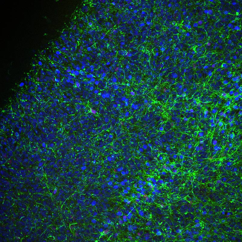
GBM-space: spatial genomic atlas of glioblastoma
About Glioblastoma multiforme (GBM)
Glioblastoma multiforme (GBM) is an aggressive type of brain tumour that is currently incurable. It is the most common type of primary malignant brain tumour in adults. The tumours are very varied – they contain a mixture of different cell types, and there is high heterogeneity in the genetic, transcriptional and epigenetic activity of the cells. In addition, different states are highly dynamic in GBM, with cells transitioning from one state to another throughout tumour development as well as in disease relapse after therapy.
The GBM-space: spatial genomic atlas of glioblastoma Project
The project is part of the Wellcome Leap ‘Delta Tissue’ or ‘tissue time machine’ project. It will use high throughput spatial genomic techniques to profile the state of cells and tissues to unprecedented detail about the states of these cancer cells and their tissue microenvironment. It is hoped that this information will help to uncover new targets for treatment development.
The collaboration draws together researchers at the Wellcome Sanger Institute, the University of Cambridge, the German Cancer Research Centre (DKFZ) and The Francis Crick Institute. It will characterise and catalogue cell type, developmental trajectory, interactions, features, form and functions to enable prediction of the transition between states.
Approach
To understand the forces that drive the cell changes seen in GBM tumours, the team comprehensively characterise the cells and molecules of the tumour tissue in 2D and 3D.
Sanger Institute scientists use spatial genomic methods to can map the intracellular signals and interactions that influence the tumour cells. The initial focus is to understand immune cells and their role in GBM by profiling neurosurgery samples from 50 patients receiving treatment at Addenbrooke’s Hospital in Cambridge.
Positions available
Computational Postdoctoral Fellow in GBM Spatial Multi-omics:
https://jobs.sanger.ac.uk/vacancy/computational-postdoctoral-fellow-in-gbm-spatial-multiomics-464137.html
Postdoctoral Fellow in GBM Spatial Multi-omics:
https://jobs.sanger.ac.uk/vacancy/postdoctoral-fellow-in-gbm-spatial-multiomics-464139.html
Staff scientist in GBM Spatial Multi-omics:
https://jobs.sanger.ac.uk/vacancy/staff-scientist-in-gbm-spatial-multiomics-464132.html
Sanger people

Dr Omer Bayraktar
Group Leader

Prof David Rowitch
Head of Paediatrics at the University of Cambridge and Honorary Faculty at the Wellcome Sanger Institute

Dr Oliver Stegle
Associate Faculty in the Cellular Genomics Programme

Sam Behjati
Group Leader & Wellcome Senior Research Fellow

Oliver Gould
PhD Student
External Contributors

Professor James Briscoe
The Francis Crick Institute

Mr Richard Mair
Cambridge University Hospitals

Adam Young
Cambridge University Hospitals

Dr Harry Bulstrode
Cambridge University Hospitals and The Francis Crick Institute

Professor David Rowitch
Department of Paediatrics, University of Cambridge

Professor Oliver Stegle
Division of Computational Genomics and Systems Genetics, DKFZ
Related groups
Affiliated Sites
External