Uncovering rules for aggressive brain tumours could lead to better treatments
Researchers at the Wellcome Sanger Institute, the European Molecular Biology Laboratory (EMBL), the German Cancer Research Center (DKFZ), the Cancer Research UK Cambridge Institute, the Francis Crick Institute, and their collaborators, have mapped over one million cells across glioblastoma tumours. This allowed them to discover that the cancer cells organise themselves according to a consistent, predictable structural pattern across different patients.
In this new study, published today (4 November) in Nature, the team was able to categorise cancer cells at different stages of their journey within the different environments in glioblastoma tumours. They looked at different time stages and used lab-grown mini tumours to mimic this development.
Along with challenging previously held beliefs about brain tumours, this roadmap opens new paths for developing targeted treatments. As the cancer cells move along in a predictable way during tumour progression, future drugs could specifically target the early developmental-like cells before they transform into harder-to-reach, resistant states.
This work has built a new foundational resource for studies to explore the mechanisms and biology of cancerous brain cells. For example, using the same single-cell multi-omic data, generated jointly for both studies, researchers also decoded the gene regulators that drive and restrict glioblastoma cell state transitions, highlighting potential targets that could help avoid treatment resistance. These results are published today in a companion paper in Nature1. Together, these papers unify understanding of the architecture of glioblastomas with insights about cancer cell plasticity, giving a holistic view of this cancer type.
Glioblastoma is an aggressive form of brain cancer. It is the most common type of brain cancer in adults, with around 3,200 people being diagnosed every year in the UK. The average survival time for people with glioblastoma is 12 to 18 months, with only 4 per cent of those diagnosed surviving five years or more2. When possible, the most common glioblastoma treatment is surgery, followed by a combination of chemotherapy and radiotherapy3.
These tumours are difficult to treat due to their location and as many tumours invade multiple parts of the brain, which can make surgery difficult or impossible in some cases.
Heidi Halls, from West Sussex, lost her dad, Steve Grant, to glioblastoma in February last year (2025).
“When Dad was diagnosed, we were told treatment wasn’t an option and that he had just 12 weeks to live. We were left with so many questions: why had this happened to him? What was happening inside his brain? What would change next, and what would those changes mean? Watching him lose his memory, hallucinate and struggle with everyday things was utterly devastating. We needed help to understand what he was experiencing and how best to support him. Even now, I am still trying to make sense of what happened. Research is urgently needed to understand this disease and develop better treatments. But families also need clear explanations, compassionate guidance and support throughout. When treatment isn’t possible, that need is still there. Being told there is nothing that can be done to treat the tumour must never mean there is nothing more that can be done for the person and their family.”
Heidi Halls speaking about her dad Steve, pictured left

How can research help?
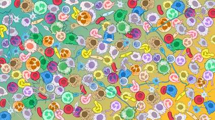
Glioblastoma treatment can also be difficult as these tumours are made up of multiple, highly dynamic cancer cell states that previously did not seem to follow any consistent rules or patterns. This means that treatment might not destroy every cell, leading to drug resistance and cancer re-emergence.
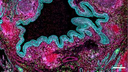
In a new study, Sanger Institute researchers and their collaborators used state of the art multi-omic technologies and tailored machine learning computational tools to map multiple sections of brain tumour samples from 12 glioblastoma patients4, combining single-cell and spatial atlassing, imaging, and whole-genome sequencing data. This range of information gives a complete understanding of each cancer cell’s molecular state, where it is in the tumour, how it is likely to change over time, how it interacts with the environment around it, and its genetic information. Additionally, the team used samples from multiple regions of the brain tumours, to give a full picture of the cells in each tumour section and how they related to each other. The resulting GBM-Space atlas (GBM meaning glioblastoma) is freely accessible.
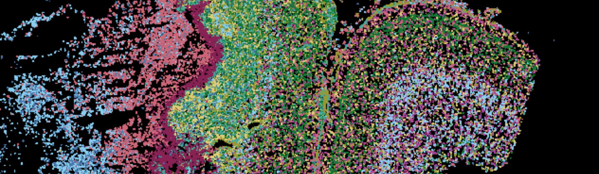
This allowed the team to see that glioblastoma cells move through the same structural pattern across each patient, following the same rules as the tumour grows:
- Cancer cells in early stages resemble brain stem cells. These developmental-like cells are found at the outer edge of the tumour, known as the leading edge, where the glioblastoma cells start to invade the surrounding healthy brain tissue. These cells act as a pool giving rise to other cancer cell states.
- As the tumour expands, these cells transform into injury response cell states, mimicking cell pathways that are involved in brain tissue repair and wound healing. These injury response cells create an inner structural layer in the tumours.
- In the later stages of brain tumour development, the core of the tumour is cut off from oxygen, meaning that all cell types transition from injury response into severely hypoxic (oxygen-deprived) states.
As well as mapping brain tumour samples, the team developed 3D models of the brain in a dish, known as brain organoids, to validate this tumour roadmap.
This research also found that each cancer cell progression stage requires specific immune microenvironments in the tumours. The team suggests that finding ways to interrupt or disrupt these environments could lead to new ways to treat aggressive brain tumours. In the future, new therapeutics could also focus on the gene regulation networks, potentially stopping cell progression, or target the early developmental cells that cannot be easily removed by surgery and often lead to cancer recurrence.
“For too long, we have approached glioblastoma treatment knowing that each patient’s tumour contains a complex mixture of cancer cells, but without a clear understanding of how best to target them all. This research gives us an opportunity to think differently. If we can anticipate the different biological states a tumour is likely to adopt, we can begin to develop treatments that target multiple aspects of the disease from the very beginning. This could fundamentally change how we design new therapies for glioblastoma, moving towards treatments that are informed not just by what a tumour looks like today, but by what it is likely to become.”
Dr Richard Mair, co-author at the Cancer Research UK Cambridge Institute and Cambridge University Hospitals NHS Foundation Trust
“Combining unbiased single-cell and multi-omics techniques with tailored computational tools allowed us to go back to the drawing board and challenge long-held assumptions about glioblastoma. We demonstrate that complex but predictable molecular networks govern cancer cell development. This work shows how computational biology can turn massive datasets into meaningful discoveries for patient care.”
Dr Oliver Stegle, co-senior author of both studies at the European Molecular Biology Laboratory (EMBL) and the German Cancer Research Center (DKFZ)
“Glioblastoma has evaded understanding for years, despite being one of the most studied types of cancer tumour. By building the largest, multi-modal atlas of these tumours to date, we have shown that these tumours are not as chaotic as previously accepted. Instead, they follow a set of rules that can be used to rewrite our understanding of glioblastoma and act as a compass to guide much needed new treatments in the future.”
Dr Omer Bayraktar, co-senior author of both studies at the Wellcome Sanger Institute and Professor at the UCL Cancer Institute
More information
- M. Saraswat, L. Rueda-Gensini, E. Heinzelmann, et al. (2026). ‘Decoding the regulatory programs governing glioblastoma plasticity’. Nature. DOI: 1038/s41586-026-11177-0
- Glioblastoma Awareness Week. Brain Tumour Research. Available at: https://braintumourresearch.org/pages/glioblastoma-awareness-week [Accessed September 2026]
- Glioblastoma treatment. The Brain Tumour Charity. Available at: https://www.thebraintumourcharity.org/brain-tumour-diagnosis-treatment/types-of-brain-tumour-adult/glioblastoma/treating-glioblastoma/ [Accessed October 2026]
- All 12 tumour samples analysed came from patients undergoing neurosurgery as part of their medical treatment. All patient surgeries were performed at Cambridge University Hospitals NHS Foundation Trust in Cambridge under approved ethical protocols with pre-operative informed consent.
The GBM-Space atlas is a freely accessible and interactive data resource, available at https://www.gbmspace.org/
Publication:
G. de Jong, F. Memi, T. Gracia, et al. (2026). ‘A spatiotemporal cancer cell trajectory underlies glioblastoma heterogeneity’. Nature. DOI: 10.1038/s41586-026-11234-8
Companion paper: M. Saraswat, L. Rueda-Gensini, E. Heinzelmann, et al. (2026). ‘Decoding the regulatory programs governing glioblastoma plasticity’. Nature. DOI: 10.1038/s41586-026-11177-0
Funding:
This research was part funded by Wellcome Leap and Wellcome. A full funding acknowledgement list can be found in the publication.